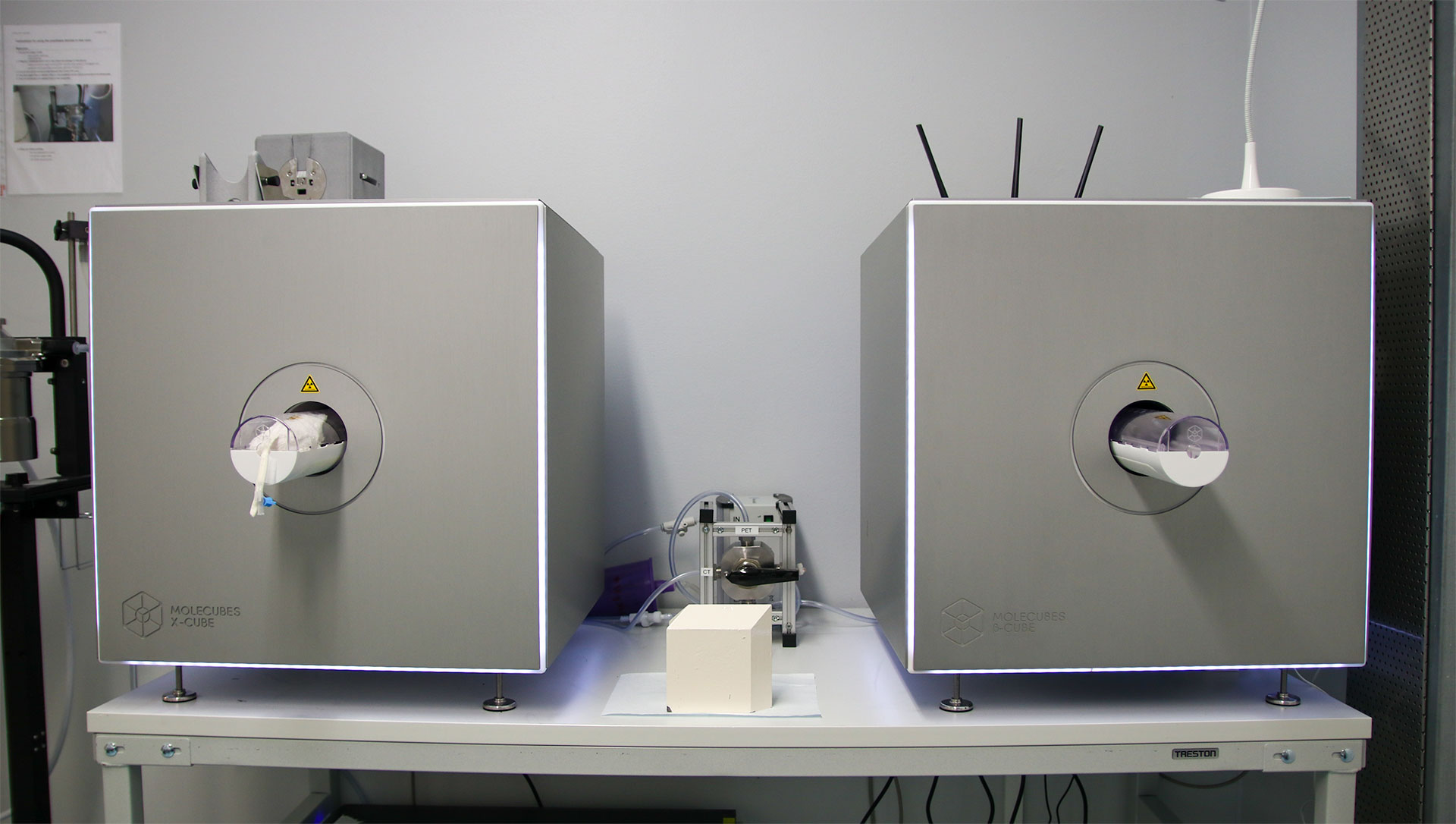
PET is non-invasive and quantitative imaging modality using molecules labelled with positron-emitting radioisotopes in tracer quantities (i.e. without pharmacological effect) to visualize and measure rates of biochemical processes (e.g. enzyme reactions, ligand-receptor interactions, cellular metabolism, cell proliferation, gene expression) in tissues of living subjects. Major advantage of PET is that the imaging procedure can be repeatedly performed, which significantly reduces the numbers of animals required for a study as compared with methods applying ex vivo measurements. The entire whole-body distribution kinetics can be determined in a single imaging study in a single animal. The same animal can be studied by PET before and after interventions, thereby allowing each animal to be used as its own control. PET imaging generally requires intravenous administration of radioactivities of the order of 0.1-5 mCi (37-185 MBq) and image acquisition times of 10-20 minutes per animal. However, isoflurane anesthesia system also allows even up to 4 hour dynamic imaging. Reconstruction of raw data into 3-D tomographical images requires several hours, and can be performed in separate workstation.
All short-lived PET tracers are synthesized by specialized radiochemists at the Radiopharmaceutical Chemistry Laboratory of Turku PET Centre.
Other supporting techniques available at PET Centre:
- Ex vivo analysis of tissues (gamma counting, digital autoradiography)
- Metabolite quantification of tissue homogenates and biological fluids
PET imaging services are provided jointly with Turku PET Centre (www.pet.fi).